
Cutting-edge Technology at Duna Medical Center
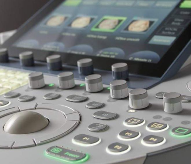
Equipped with the latest devices, our radiology department not only features a low-dose X-ray machine but also a specially equipped 3D tomosynthesis-capable Hologic digital mammography device suitable for stereotactic tissue sampling, and a whole-body bone densitometer, all aiding in precise diagnosis.
3D, tomosynthesis-guided stereotactic breast biopsy: A specialized tool for investigating microcalcifications, potentially avoiding many breast surgeries.
The stereotactic vacuum-assisted breast biopsy is recognized worldwide as the only perfectly accurate procedure for the non-surgical clarification of breast microcalcifications and appropriate surgical planning. The most modern method for investigating changes found during screening and clinical mammography examinations, which are not visible on ultrasound and not palpable, is tomosynthesis-guided stereotactic sampling, uniquely available only at Duna Medical Center in our country.
Its great advantage is its 99.3% accuracy, meaning that breast surgery is practically only performed in the case of malignant changes. This is particularly important because without an accurate diagnosis, two-thirds of "blindly" operated microcalcifications turn out to be benign, so with such sampling, breast surgery can be avoided in the vast majority of cases involving microcalcifications.
Digital mammography with 3D tomosynthesis: 41 percent more breast cancer can be detected.
The special, tomosynthesis-equipped direct digital mammography device at Duna Medical Center rotates around the breast in an arc, separating the complex mammography image into about 50 slices, each 1 millimeter thick. (Most traditional - even digital - mammography devices used elsewhere take only a single image from one direction.) Our device's specially sensitive, newly developed detector operates at a very low dose and provides very high-resolution images - the smallest image point is just 0.07 millimeters.
As a result of tomosynthesis, scientifically proven and described in the literature:
- 29-41 percent more invasive breast cancers can be detected than with traditional digital mammography,
- more breast cancers are found earlier and at a smaller size, allowing treatment to start sooner,
- 40 percent fewer additional images need to be taken,
Our Hologic device is unique in the world for its ability to generate a summed-up - so-called synthetic - 2D mammography image from tomosynthesis layer images without X-rays, which has been accepted by the American FDA.
To analyze the results of such complex imaging examinations and perform special biopsy procedures, highly trained professionals are needed: Duna Medical Center employs specially trained radiologists with international practice and breast imaging certification.
Automatic X-ray image fusion for examining orthopedic abnormalities and scoliosis
With our special device, whose software automatically stitches together multiple series X-ray images from a long area (such as all sections of the entire spine), the spinal column can be depicted on a single digital panorama image. This allows for a good examination of spinal statics, accurate measurement of the degree of scoliosis, and precise calculation of arcs, angles, and distances. This significantly aids in determining the treatment methods for orthopedic deviations.